
What Is Age-Related Macular Degeneration (AMD) and How Is It Treated?
Physical Health
Macular degeneration is an eye disease that can cause vision loss. Since it’s more common in older adults, it’s often referred to as age-related macular degeneration, or AMD. AMD is a very common condition and one of the leading causes of vision loss. It’s estimated that about 20 million adults in the United States (U.S.) are living with AMD. Continue reading to learn more about AMD, how it’s diagnosed, and treated.
What Is AMD?
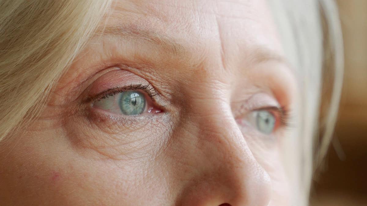
AMD is a progressive eye disease that can make your central vision blurry. Over time, AMD can also lead to central vision loss. Your central vision is the part of your vision you use to see straight ahead of you. Most people with AMD-related vision loss aren’t completely blind because AMD doesn’t affect your peripheral vision (what you see out of the corner of your eye). That means if you have AMD and look at the center of a clock, you’ll be able to see the numbers around the outside of the clock, but not the hands.
AMD affects central vision because it’s caused by damage to the macula — the center part of the retina. The retina is the tissue at the back of the eye that senses light and converts it into nerve signals. These nerve signals are transmitted to the brain by the optic nerve. When the macula is damaged, the brain may have difficulty translating the nerve signals to an image, making vision appear blurry.
The cause of AMD depends on the type.
Dry vs Wet AMD
There are two main types of AMD — dry AMD and wet AMD.
About 90% of people diagnosed with AMD have dry AMD. In dry AMD, the macula is damaged by abnormal deposits of fats and proteins, called drusen. It’s normal for older adults to have a few small drusen. However, many drusen or large drusen are a sign of AMD. Large drusen can build up on the macula, making it thinner.
About 10% of people with dry AMD can develop wet AMD, a more serious form of AMD. In wet AMD, abnormal blood vessels form under the macula. Wet AMD is also called neovascular AMD since this is the medical term for new blood vessels. These new blood vessels can leak blood and other fluids into the macula, causing damage and scarring. Wet AMD typically causes vision loss faster than dry AMD.
AMD Stages
AMD is classified into three stages, based on severity.
In early AMD, some drusen can be found on the macula. In this stage, the drusen don’t usually affect vision or cause any symptoms. Over time, some people may develop larger drusen that cause some symptoms and vision changes. This is known as intermediate AMD.
Late AMD is the most advanced stage of AMD. In dry AMD, it’s known as geographic atrophy. In this stage, large patches of the macula appear atrophied (thinner), causing more severe symptoms. Wet AMD is always considered late AMD because it can cause severe vision loss more rapidly than dry AMD.
You may have AMD in one or both eyes. It’s possible to for each eye to be in a different stage of AMD. Additionally, you can have wet and dry AMD in the same eye.
Who Gets AMD?
Most people with AMD are older than 50. As the name suggests, age-related macular degeneration is more common with age. People older than 75 years are three times more likely to develop AMD compared to people aged 65 to 74 years.
Additional risk factors for AMD include:
- Eating foods high in saturated fat
- Overweight and obesity
- High blood pressure
- High cholesterol
- Heart disease
- Family history of AMD
- Smoking
What Are the Symptoms of AMD?
In early AMD, the symptoms can be subtle, and many people don’t notice any vision changes. It may be even harder to notice AMD symptoms if only one eye is affected. Some of the early signs of AMD may include needing brighter lights to read and more difficulty seeing at night.
AMD is a progressive disease. That means that, without treatment, AMD worsens with time. In intermediate AMD, some people may begin to notice some symptoms, such as:
- Blurry vision
- Difficulty seeing in low light
- Colors that don’t seem as bright or vivid as they used to
- Blank spots in the field of vision (called scotoma)
As AMD progresses, the area of blurriness may get larger and more blurry. Some people may also notice more or larger blank spots that appear in the field of vision. It may become more difficult to read or recognize faces.
Some people with late AMD have vision changes that make straight lines appear wavy or crooked. This symptom can be a warning sign of serious vision problems. If you notice that objects appear curved when you know they should be straight, it’s important to seek medical care right away to prevent permanent vision loss.
How Is AMD Diagnosed?
AMD is diagnosed by an ophthalmologist — a doctor who specializes in diagnosing and treating eye diseases. Ophthalmologists conduct a comprehensive eye exam to detect the signs and symptoms of AMD. There are several different tests involved in a comprehensive eye exam. We’ll discuss them below.
Eye Exam
An eye exam consists of several tests to check your vision, including:
- Visual acuity test — to test how clearly you can read letters from close or far
- Visual field test — to check your peripheral vision
- Eye muscle function test — to check how well you’re able to follow an object with just your eyes
- Tonometry test — to check the pressure inside your eye
- Amsler grid test — a test where you look at a grid of straight lines to check if you see any distortions or wavy lines
Dilated Eye Exam
A dilated eye exam is an exam where an ophthalmologist instills eye drops that dilate (widen) your pupil. Dilating the pupil lets in more light and makes it easier to see the back of the eye to look for problems. Once the pupil is dilated, your ophthalmologist will look inside your eye with a special lens. They’ll look for signs of drusen, geographic atrophy, and abnormal blood vessel growth.
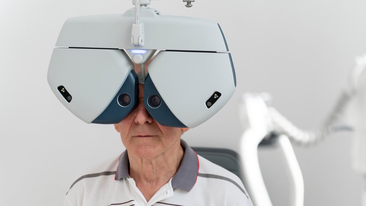
Imaging Tests
Imaging tests for AMD use special cameras to take pictures of the back of your eye. An optical coherence tomography (OCT) test uses a special machine to take pictures of the back of your eye. If your ophthalmologist suspects you may have wet AMD, you may need an optical coherence tomography angiography (OCTA). This test uses a laser to check the blood flow in the eyes.
A fluorescein angiography is another test to check for wet AMD. In this test, you’ll be injected with a special dye called fluorescein. When the dye flows through the blood vessels in the eye, it can help identify areas where blood is leaking into the macula.
Is There a Cure for AMD?
There isn’t a cure for AMD. However, early diagnosis and treatment can help slow the progression of AMD and help prevent vision loss. Your treatment depends on the type of AMD you have.
How Is Dry AMD Treated?
AMD treatment depends on what stage of AMD you have. Currently, there isn’t an effective medical treatment for early AMD. If you have early AMD, your ophthalmologist will closely monitor you for signs that AMD is progressing to intermediate dry AMD.
Intermediate Dry AMD Treatment
If you have intermediate dry AMD, your ophthalmologist may recommend you take a supplement that’s specifically designed for people with AMD, called the AREDS 2 supplement. Clinical trials have found that the AREDS 2 supplement can prevent people with intermediate dry AMD from progressing to late AMD.
The AREDS 2 supplement contains a unique mix of several ingredients, including:
- Vitamin C
- Vitamin E
- Copper
- Zinc
- Lutein
- Zeaxanthin
While many of the ingredients found in the AREDS 2 supplement can be found in food and other supplements, AREDS 2 supplements have the right amount and combination of ingredients proven to help AMD.
The large amounts of vitamins and minerals in the AREDS 2 supplement can cause some side effects. For example, it can affect how your body digests food and how well other medications work. Additionally, the first version of the AREDS supplement contained beta-carotene. This ingredient has been found to increase the risk of lung cancer in people who smoke or people who used to smoke.
Although the AREDS 2 supplement is available without a prescription, it's important to talk to your physician before you start taking this supplement.
Geographic Atrophy Treatment
Treatment options for people with geographic atrophy include medication and surgery.
Medications for geographic atrophy are given by an intraocular (inside the eye) injection once every one to two months. Pegcetacoplan (Syofovre) and avacincaptad pegol (Izervay) are examples of intraocular injections for geographic atrophy. These medications may be able to help slow the progression of late dry AMD but can’t help you recover vision loss that’s already occurred.
Surgery for geographic atrophy involves replace the lens of your eye with an implantable miniature telescope (IMT). The IMT helps to magnify what you see in your central vision and focus light on undamaged parts of the retina.
How Is Wet AMD Treated?
Treatments for wet AMD aim to stop the growth of new blood vessels.
As in geographic atrophy treatment, medications for wet AMD are also given by an intraocular injection. These medications help prevent more damage from leaky blood vessels by blocking a protein that encourages the growth of new blood vessels, called vascular endothelial growth factor (VEGF) protein. By blocking VEGF, these medications prevent the formation of new blood vessels in the macula. Examples of medications for wet AMD include:
- Aflibercept (Eylea)
- Faricimab (Vabysmo)
- Bevacizumab (Avastin)
- Ranibizumab (Lucentis)
Less common treatments for wet AMD include laser treatments and photodynamic therapy. Laser treatments are another treatment option that can help stop the growth of new blood vessels in the macula. Photodynamic therapy is a treatment that uses a laser to target abnormal blood vessels in the eye using an injectable drug that’s light sensitive.
What Are the Side Effects of AMD Treatment?
Each AMD treatment has the potential to cause side effects. Possible side effects and complications of AMD treatments include:
- Eye infections
- Retinal detachment (when the retina separates from the back of the eye)
- Cataracts
- Vision loss
Can You Prevent AMD?
There’s no sure way to prevent AMD. However, there are some things you can do to lower your risk factors. If you already have AMD, following these tips may help you manage your symptoms and prevent AMD progression.
Maintain Healthy Lifestyle Habits
Healthy lifestyle habits may help prevent the development or progression of AMD, including:
- Don’t use tobacco products
- Eat a balanced diet rich in fruits and vegetables (especially green leafy vegetables, like spinach and kale)
- Get regular exercise
- Maintain a healthy weight
- Wear sunglasses to protect your eyes from the sun

Get Regular Eye Exams
Regular eye exams can help detect eye diseases, like AMD, before they begin causing symptoms. When AMD is caught early, there are more options available to help slow the progression.
It’s recommended that everyone over the age of 60 get a dilated eye exam every one to two years. You may need a dilated eye exam more frequently if you have diabetes, high blood pressure, or a family history of eye disease.
Monitor For Vision Changes at Home
If you’ve been diagnosed with any type or stage of AMD, an eye care professional may recommend using an Amsler grid every day. Regular use of this simple tool can help you detect serious vision problems in an early stage.
The Amsler grid can help you identify subtle vision changes that may not be easy to detect in daily life. Your ophthalmologist can teach you how to use the Amsler grid. If you notice any changes in how the Amsler grid looks from one day to the next, it’s important to contact your physician right away to prevent permanent vision loss.
Manage Other Health Conditions
Having other health conditions can increase your risk of AMD. For example, overweight and obesity, high blood pressure, and high cholesterol all increase the risk of developing AMD. Work with your physician to manage these health conditions to reduce your risk of vision problems from AMD.


